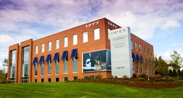
OPTX Rhode Island is well recognized for its commitment to technology, allowing us to gather an extraordinary collection of information about a patient’s eyes and vision. For example, we have imaging devices that identify eye disease states at the cellular level. Additionally, we have the latest technology to assist us in dispensing eyeglass and contact lens prescriptions with extraordinary accuracy. The result of these impressive technologies is earlier diagnosis of disease and more accurate assessments, all leading to better outcomes for our patients. Quality resonates throughout our practice and we are proud of it.
- EPIC 5100 refraction system
- OPD scan III refractive power/corneal analyzer
- Nikon speedy-k autorefractor
- Pentacam corneal topography
- Corneal pachymetry
- Tomey specular microscopy
- Haag-Streit slit lamps
- Anterior segment digital photography
- OPTOS retinal imaging system
- Zeiss Visucam fluorescein angiography system
- Heidelberg spectralis optical coherence tomography
- B-scan ophthalmic ultrasonography
- Humphrey visual field analyzer
- Diopsis ERG and VEP
- Ocular Response Analyzer
- Foresee PHP- preferential hyperacuity perimeter
- Lenstar
- Immersion a-scan ultrasound
- LUMA eyemagination
- TearLab Osmolarity System
- RPS InflammaDry Detector
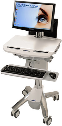
 EPIC 5100 refraction system
The Epic-5100 is a highly accurate digital refraction system. Many of the tests that were previously very time-consuming during a typical eye exam are now performed with greater precision using this technology. The end result is a better assessment of your vision needs. Best of all, the process is completed quickly and easily.
The Epic-5100 refraction system integrates wavefront data into the comprehensive eye exam allowing us to better understand many patient visual complaints that were difficult to assess in the past. We have developed a specific testing protocol with the Epic 5100 that allows us to deliver the best possible eyeglass and contact lens prescription, truly customized for each patient.
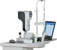
 OPD scan III refractive power/corneal analyzer
The OPD (Optical Path Difference) is essential in determining low and high order aberrations, identifying astigmatism, surgical planning for toric and multifocal intraocular lenses in cataract surgery, accurately recording pupillary size, aligning proper centration of intraocular lenses, delivering wavefront-optimized eyeglass prescriptions as well as separate, specific day and night eyeglass prescriptions. The data we obtain from this technology enables us to prescribe an individualized treatment plan for a variety of vision problems.
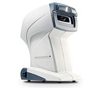
 Nikon speedy-k autorefractor system
The Speedy-K autorefractor is another diagnostic tool used in assessing the visual system to refine eyeglass prescriptions.
 Pentacam corneal topography
Pentacam is the gold standard for evaluating the cornea. It actually measures 25,000 true elevation points of the cornea, capturing dramatically enhanced, precise image representation. These data points are then used to generate maps for diagnosis and treatment of a variety of corneal diseases. Dr. Diamante is one of the few ophthalmologists in New England to have this technology in his practice.
 Corneal pachymetry
Corneal pachymetry is a test used to determine thickness of the cornea. Our practice utilizes a corneal contact ultrasound as well as a non-contact method with the Pentacam system. Corneal pachymetry is essential when planning for corneal astigmatism procedures as well as detecting early glaucoma. The thickness of the cornea can predict glaucoma development when combined with measurement of intraocular pressure.
 Tomey specular microscopy
Tomey Specular microscopy is a non-invasive photographic technique that facilitates rapid and accurate diagnosis of corneal endothelial disease. The corneal endothelium is responsible for maintaining corneal clarity. Corneal endothelial disease can lead to edema, loss of transparency of the cornea, and ultimately loss of vision. With this technology, we are able to accurately measure structure and function of the corneal endothelium.
 Haag-Streit diagnostic slit lamps
Our practice provides our doctors with the highest quality standards in eye exam equipment. Haag-Streit slit lamps provide exceptional detail in evaluating eye disease. This greatly assists our doctors to ensure the most accurate diagnoses from the cornea to the retina.
Back to top
 Anterior segment digital photography
Our practice has an advanced digital slit lamp camera that allows us to view and archive pathology of the front part of the eye, also known as the anterior segment. The anatomy of the anterior segment includes the eyelids, sclera, conjunctiva, cornea, iris, pupils and lens. Because of this technology, our doctors are able to better document and follow disease progression of the anterior segment.
 OPTOS retinal imaging system
OPTOSThe Optos technology redefines new standards in retinal imaging with ultra-high definition detail. This simultaneous pole to peripheral view of the retina allows us to diagnose, analyze and monitor ocular pathology that may first present in the periphery of the retina. More than 80% of the retina is imaged in a single view. The typical examination of the retina is performed in serial sections, allowing for partial viewing each time. Optos provides simultaneous evaluation of the central and peripheral retina allowing for much easier assessment of retinal conditions.
 Zeiss Visucam fluorescein angiography system
Zeiss Viscam Fluorescein angiography is a technique used for examining blood vessel circulation of the retina. This is accomplished by using a fluorescent dye and a specialized digital fundus camera. The Zeiss system provides high-resolution 3D imaging for optimized visualization of critical retinal and choroidal blood vessels. This detail gives us confidence in diagnosing subtle, challenging retinal and choroidal disease.
 Heidelberg spectralis optical coherence tomography
The Heidelberg OCT assists in detecting microscopic structural changes of the macula and optic nerve head in advance of vision loss. This allows for earlier treatment of devastating diseases such as age-related macular degeneration, diabetic retinopathy and glaucoma. These 3D images have revolutionized diagnosis and management of these common eye diseases.
 B-scan ophthalmic ultrasonography
B-scan ultrasound is useful in direct visualization of intraocular structures when the typical view is difficult or impossible. This occurs in cases such as severe corneal swelling, densed corneal scars, bleeding in the front part of the eye (hyphema), dense, cloudy cataracts or bleeding in the back of the eye (vitreous). B-Scan ultrasonography is useful in differentiating iris and ciliary body lesions, characterizing intraocular tumors, as well as better defining retinal detachments and disc drusen versus disc edema.
 Humphrey visual field analyzer
Analysis of the visual field can detect dysfunction in central and peripheral vision which can be caused by various medical conditions such as glaucoma, stroke, brain tumors and other neurologic deficits. This computerized test maps and calculates a patient’s field of vision. It reveals a wealth of information regarding the health of a patient’s visual system.
 Diopsis ERG and VEP
The Diopsis Electroretinograph (ERG) tests the performance of the inner retinal cells of the eye, especially useful for earlier detection for more specific and timely management of glaucoma and diseases of the retina, including macula degeneration in diabetic retinopathy.
The Diopsis Visual Evoked Potential (VEP) measures electrical activity in the vision system.The VEP helps to determine how your eyes communicate with your brain. It can help detect mechanical or neurologic abnormalities related to vision, which are often subtle and difficult to detect.
 Ocular Response Analyzer
This device is the only device that measures Corneal Hysteresis, a superior predictor of glaucoma progression.
 Foresee PHP- preferential hyperacuity perimeter
The Foresee PHP provides an unsurpassed level of sensitivity in detecting macular degenerative changes. This visual field analyzer specifically monitors macular function. Macular visual field changes can be secondary to development of wet age-related macular degeneration.
 Lenstar
Lenstar employs optical coherence interferometry. It quickly performs accurate axial length and keratometry readings which are critical components in calculating the correct intraocular lens power in cataract surgery. The accuracy of this technology and its integration with the most sophisticated intraocular lens calculation formulas, allows Dr. Diamante to deliver ultimate precision to your cataract surgery outcomes.
 Immersion a-scan ultrasound
Immersion a-scan ultrasound is used to determine the axial length of an eye when optical coherence interferometry is not possible. There are types of cataracts, as well as certain eye lengths, that require this technique to ensure accurate calculation of the intraocular lens power for cataract surgery.
 LUMA eyemagination
Luma Eyemagination software is an interactive tool which provides a clear, 3D animation of eye disease as well as treatment options. The software imagery allows patients to better comprehend disease entities and treatment options available to them. Additionally, LUMA can help patients understand what happens when an eye condition progresses and leads to vision problems.
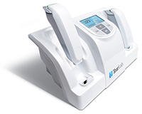 TearLab Osmolarity System
The TearLab Osmolarity System is intended to measure the osmolarity of human tears to aid in the diagnosis of dry eye disease in patients suspected of having dry eye disease, in conjunction with other testing methods.
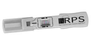 RPS InflammaDry Detector
The RPS InflammaDry Detector is the first and only rapid, point-of-care test to detect for MMP-9, an inflammatory marker that has consistently been shown to be elevated in the tears of patients with Dry Eye disease.
Trust World-Class Skill and Technology
For exceptional eye care and treatment using the latest technology and equipment, come to Giulio Diamante, MD & Associates here at OPTX Rhode Island. Call us at 401.521.3606 or use our online Request an Appointment form to schedule your appointment.